July 18, 2024 feature
This article has been reviewed according to Science X's editorial process and policies. Editors have highlighted the following attributes while ensuring the content's credibility:
fact-checked
peer-reviewed publication
trusted source
proofread
Exploring how astrocytes respond to spinal cord injury or stroke-induced tissue damage

Past neuroscience studies found that when the central nervous system (CNS) is damaged, for instance following a stroke or spinal cord injuries, the lesions become surrounded by borders of newly proliferated astrocytes.
Astrocytes are star-shaped glial cells known to have a range of functions, ranging from clearing neurotransmitters to promoting the formation of synapses in healthy brains to responding to injury and disease.
While the proliferation of reactive astrocytes around damaged CNS tissue is well-documented, the unique features of these cells and their contributions to wound repair have not yet been uncovered. Better understanding of astrocytes around lesion borders could inform the development of more effective and targeted therapeutic strategies to promote recovery after injuries or strokes.
Researchers at University of California, Los Angeles recently carried out a study aimed at better understanding the responses of astrocytes to CNS tissue damage. Their findings published in Nature Neuroscience, suggest that after adult mice suffer spinal cord injuries or strokes, 90% of the border-forming astrocytes around their lesions are derived from proliferating local astrocytes, while 10% are derived from oligodendrocyte progenitor cells.
"In addition to nerve cells, the brain and spinal cord contain other cell types that are important for support functions and responding to injury, infection and disease," Michael Sofroniew, co-author of the paper, told Medical Xpress. "Astrocytes are one such cell type. Until recently (the last 25 years) these other cell types, including astrocytes, were largely ignored and their roles were poorly understood."
In the past, astrocytes were thought to prevent nerve cells from regrowing after injuries or strokes. This previous belief was what initially inspired Sofroniew and his colleagues to investigate these cells, yet their studies found that it was far from a reality.
In fact, two of their previous papers published in Nature in 2016 and 2018, outlined experimental evidence that suggested the opposite is true. Instead of preventing the regrowth of damaged nerve cells, the team found that astrocytes could support cell regrowth and lesion repair following injuries.
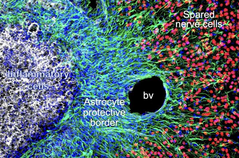
"The current study follows on from this earlier work and examines in detail why and how astrocytes respond to tissue damage," Sofroniew explained. "We show that in response to stroke or traumatic injury, astrocytes multiply and form neuroprotective borders around damaged areas.
"These newly generated astrocyte borders serve to protect immediately neighboring spared neurons by surrounding the inflammatory cells that infiltrate into the damaged area, to clean up debris and protect against infection."
The borders formed around lesions essentially enclose inflammatory cells, preventing them from spreading and damaging spared nerve cells. The astrocytes forming these borders are thus now referred to as 'wound repair astrocytes'.
As part of their recent study, Sofroniew and his colleagues set out to investigate how astrocytes in the brain and spinal cord of adult mice respond to tissue injuries. To do this, they used a combination of techniques, which allowed them to run histological tissue sections and biochemical gene expression analyses.
"For many years, astrocytes were regarded as detrimental cells after traumatic injury, stroke or disease," Sofroniew said. "In contrast, our findings show that astrocytes exert neuroprotective functions in many disorder contexts. There are emerging hints that some of these functions become less effective as aging progresses or in certain disease contexts."
The results gathered by the researchers suggest that after an adult mouse suffers a stroke or CNS injury, local mature astrocytes de-differentiate, proliferate and become genetically reprogrammed to serve specific functions.
As a result of these processes, molecules associated with astrocyte-neuron interactions are downregulated, while molecules that interact with immune cells and associated with wound healing are upregulated.
These observations contribute to the understanding of astrocytes and their role in the repair of CNS lesions. In the future, they could pave the way for further studies focusing on the features of wound repair astrocytes, potentially leading to more important discoveries.
"Understanding the basic biology of astrocyte functions in response to trauma and stroke, and ultimately understanding how these responses are regulated by molecular signals, has the potential to identify new therapeutic targets that may improve outcomes," Sofroniew added.
"Our next goal will be to identify signaling molecules that regulate wound repair by astrocytes after traumatic injury or stroke and that might be targeted to improve wound repair by sparing more functional tissue and thereby improve the outcome."
More information: Timothy M. O'Shea et al, Derivation and transcriptional reprogramming of border-forming wound repair astrocytes after spinal cord injury or stroke in mice, Nature Neuroscience (2024). DOI: 10.1038/s41593-024-01684-6.
© 2024 Science X Network


